
So, you found a lump?
Nobody likes to find a lump on their body that wasn’t there the last time they checked. When a doctor visit and subsequent medical testing doesn’t provide immediate identification of the growth, a biopsy may be ordered.
What does this entail?
A biopsy is an examination of tissue or cell samples from a living body. When removed, usually surgically, the samples are sent to a laboratory to help diagnose a physical malady. Some biopsies can be performed with minimal anesthesia and “invasion” (penetration into the body) – others, not so much.

A biopsy is the only proven way to diagnose most cancers and many other diseases. In medical terms, cells or tissues can be either benign (not diseased), malignant (diseased), or infectious (diseased and capable of being spread to others).
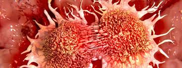
 Cancer cells
There are many different kinds of biopsies:
Skin biopsy – For a rash or lesion on the skin’s surface that doesn’t go away on its own or respond to treatment, the doctor may scrape, cut, or punch a small piece of skin to check for infection, inflammation, or cancer.
Bone marrow biopsy – As the name suggests, a long needle inserted into the hipbone is used to extract a bone marrow sample for analysis. Both cancerous and noncancerous conditions can be detected, including leukemia, anemia, infection, or lymphoma.
Endoscopic biopsy – In this procedure, a thin, flexible tube with a miniature camera and light at the end, called an endoscope, sends real-time images to a video monitor. After identifying targeted tissues, the doctor can insert small surgical tools into the endoscope to collect a sample. With insertion points at the mouth, nose, rectum, or urethra, most internal organs (e.g., lung, liver, or colon) are accessible.
Needle biopsy – When skin or subcutaneous tissue lying just under the skin needs to be collected, the doctor uses a needle specific to the task: a core needle removes a column of tissue (like a geologic core sample of layers deep inside the earth), whereas fine needle aspiration (FNA) is where a thin, hollow needle attached to a syringe draws out fluid and cells. FNA is considered one of the least invasive methods of biopsy.
Image-guided biopsy – This is a type of non-surgical needle biopsy that is assisted by X-ray, ultrasound, mammography, CT or MRI imaging. Performed when an abnormal mass or lump is found in a hard-to-reach organ (thyroid, liver, kidney, lung) or body part (bone, abdomen, pelvis, lymph nodes), radiologists can locate the right spot for sampling and avoid injuring nearby healthy tissue.
Surgical biopsy – The most invasive type of biopsy requires an actual surgical operation. This option is reserved for when none of the other methods can reach the target site inside the body or have successfully identified the cause of the problem. Using the least invasive tools available, the surgeon passes through small incisions to examine the area and perform the minor surgery.
Liquid biopsy – One of the newest, minimally invasive diagnostic procedures is also called a fluid or fluid phase biopsy. Small pieces of tumor material from bodily fluids (blood, plasma, serum or urine) are collected. The underlying premise of this diagnostic test is that tumors shed molecules and cells into bodily fluids. However, this technique is still being developed, and results are under review by healthcare professionals. The FDA approved the first liquid biopsy for cancer in June 2016.
No matter how you slice it, biopsies empower both diagnostician and patient with the power of knowledge. Identifying at-risk cells and tissues in the early stages of disease pays off with improved chances of recovery and cure.
If you notice an unusual bump or lump where there hadn’t been one before, see your doctor right away. Treatment might be just a biopsy away.

Recent Comments